Rady Children’s Hospital-San Diego has recruited a prominent surgeon-researcher within its Orthopedics & Scoliosis program to advance knowledge of the genetic and biomechanical factors behind scoliosis; advances in patient care can lead to better outcomes and quality of life.
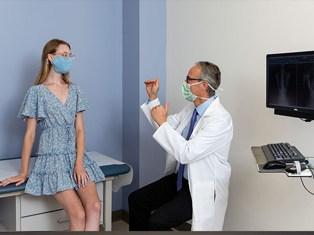
When Michael Kelly, MD, director of scoliosis and spinal deformities at Rady Children’s Hospital, is asked to explain how this spine-twisting disease affects adolescents differently from adults, he offers a blunt example.
“Not many adults ask if they’ll be able to ride a roller coaster again after surgery,” he says. “For adults, spine surgery is mostly about removing pain. For children it’s not always about pain, it’s about an issue that can be with them for their entire lives.”
The S-shaped curves characteristic of scoliosis develop most frequently during pre-pubescent growth spurts. Most instances of scoliosis are classified as idiopathic – they emerge seemingly out of nowhere, with no underlying spine or neuromuscular causes. For these reasons, pediatric research centers such as the Spine Center at Rady Children’s are emerging as powerhouses when it comes to treatment and understanding of spinal disorders.
“Before I came to Rady Children’s, I was getting pulled in every direction,” recalls Kelly, who joined after completing two spine fellowships at Washington University in St. Louis. “Being able to focus on children instead of a diverse patient mix is beneficial from a research perspective. And having the chance to work with Rady Children’s surgeon-in-chief Peter Newton, MD who’s contributed so much to this field and has such intuitive, emotional intelligence – it was an opportunity I couldn’t pass up.”
With Kelly on board, the Rady Children’s Spine Center is looking to build on its record of patient-centred innovation. But the centre also aims to advance the fundamental understanding of scoliosis. In partnership with genomic specialists, the team is investigating the genetic basis behind the condition’s development and the immunological factors that may govern patients’ recovery from corrective surgery.
Restoring balance
Newton has pioneered data-driven techniques to improve scoliosis treatments for more than 25 years at Rady Children’s. He notes that the most common method in severe curvature cases is to perform spinal fusion, an operation that transforms twisted vertebrae into a solid block of bone with the aid of bone grafts and implants such as rods and screws.
“We’ve become exceptional at correcting scoliosis using fusion technology, but it’s highly dependent on the surgeon’s understanding of three-dimensional deformities,” says Newton. “You need experience to execute these technically challenging operations, and from knowing Mike Kelly’s performance as a surgeon and researcher, this was the person I wanted to join our team.”
With more than 150 peer-reviewed articles on spine surgeries, Kelly’s research focuses on classifying and interpreting the biomechanical issues surrounding spinal surgery – identifying, for instance, how fusions that harmonize interactions between different anatomical planes and pelvic bones can play a key role in restoring a sense of balance.
“We’re putting metal in and ‘gluing’ these bones together that are supposed to move; it’s one of the most biologically abnormal things we could do,” says Kelly. “If you fuse a shoulder, fuse an elbow – every orthopedic physician knows what angle to lock in the bones even if it’s quite rare. But very few surgeons think about where, in precise three-dimensional space, spinal bones should be fused, and these are done thousands of times a year.”
Getting a grip on scoliosis
In vertebral body tethering, flexible cords are implanted along the side of the spinal column to correct scoliosis without spinal fusion.
Newton, on the other hand, is renowned for developing spinal growth tethering surgery, an innovative strategy to straighten the spine without resorting to bone fusion. In this procedure, flexible polyethylene cords are implanted asymmetrically along the side of the spinal column to influence growth, eventually straightening the spine as the patient grows.
“After doing research in this field for more than two decades, we’ve moved beyond animal studies and basic science into what happens clinically when patients have these implants,” he says. “How do we modulate the growth appropriately? And what are the factors that predict the success of an operation like this?”
Through innovative technology such as an orthopedic imaging machine that can capture biplanar full-body X-rays of the spine while patients stand upright, Newton and his team track the three-dimensional shape of the tethered vertebra over two to three years of growth. If, for example, they spot trapezoidal-shaped vertebra, they can utilize tension in the tethering cord to let the short side catch up in growth until healthier rectangular bones appear once more in the X-rays.
Newton emphasizes that the success of the tethering approach depends on finding patients with enough capacity to modulate growth. “Right now, our ability to find the perfect patient is modest at best,” he says. “If you put the tether in too late after the growth spurt, not much happens. And even then, how much you correct initially with the cord, how much additional pressure you need over time – that varies based on the patient curve size and remaining growth.
“There are some cases where it just doesn’t work as well as we’d predict, and we estimate as many as 25% of patients may end up undergoing a posterior spinal fusion within five years. That’s 75% of people where you’ve maintained nearly all the flexibility of the spine, avoiding spinal fusion. But for the patients who undergo two operations instead of one, we’ve got to get better at making predictions We’ve had some spectacular outcomes with tethering, especially in a 15-year-old patient named Sophie Allison.”
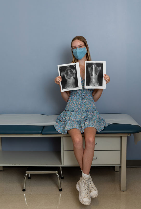
Sophie’s Story
Sophie, a healthy and active 15-year-old, was referred to Rady Children’s for an evaluation of scoliosis. Upon examination, it was found that she had a substantial shift of her trunk to the right side, with a large right thoracic rib prominence. While her neurological examination was normal, there were features of her curvature that made the doctor suspicious of an underlying neurological cause for her scoliosis.
“She had an unusually large degree of thoracic roundback, or kyphosis, which is not typical of adolescent idiopathic scoliosis (AIS),” Newton notes. “In fact, most AIS has a relative flatback or loss of normal kyphosis. Her X-ray confirmed a 60-degree long thoracic scoliosis with 65-degree thoracic kyphosis.”
Additionally, an MRI of her spine revealed a large syringomyelia of her cervical and thoracic spinal cord, along with a Chiari malformation. The presence of the cerebellar tonsil within the cervical spinal canal was the likely cause of the syringomyelia. Sophie was referred to a neurosurgical colleague, David Gonda, MD, who performed a craniectomy and C1 laminectomy to decompress the cranio-cervical junction.
Once Sophie had recovered from her Chiari decompression surgery, Newton performed scoliosis correction with a posterior instrumentation and fusion from T2-L3, achieving segmental pedicle screw fixation using a freehand technique. Screw positions were verified before corrective maneuvers began. The right and left rods were differentially contoured to impart axial plane rotational correction while reducing the kyphosis and fully correcting the scoliosis. Segmental compression and transverse plane manipulation of the screws finished the 3-dimensional correction of Sophie’s spinal deformity. Intraoperative neuromonitoring was performed throughout the procedure due to the intraspinal pathology noted on the preoperative MRI, but there were no monitoring changes during surgery.
Following the surgery, Sophie followed a multimodal pain regimen and enhanced recovery after surgery pathway and was discharged on postoperative day three. Over the following 6 months, Sophie returned to her normal activities. Now, 2.5 years after her surgery, Sophie is living her life with scoliosis in the rearview mirror.
Hidden in the genes
Using tiny bits of bone and muscle left over from spinal surgeries, Newton and his team are working with colleagues at the Rady Children’s Institute for Genomic Medicine to uncover genetic markers that may indicate scoliosis in cases like Sophie’s. Currently, he favors the theory that the mechanisms controlling bone growth become out of sync in patients with idiopathic scoliosis, creating curves and twists to accommodate extra spine length.
“Once the spine rotates a little bit off to the side, that vertebra is not in the area where it’s getting its normal weight bearing, and it starts a cycle of asymmetric growth with wedged vertebral shape that gets worse and worse,” Newton explains. “With tethering we can reverse that cycle; that’s what makes it such an elegant solution.”
Along with expanding knowledge of the genetic underpinnings of scoliosis, Kelly foresees a continuing focus on patient-centered innovations at Rady Children’s. Already experienced in data analytic techniques to determine risks and track reported outcomes, he plans to tackle two key issues: the use of immunology to improve patient self-healing, and the expansion of artificial intelligence algorithms to guide surgeons.
“If we can come up with an AI system that can analyze these deformities and design the surgery so that you’re getting the same treatment whether you’re in San Diego, or Idaho, or Tunisia – that’s what we need,” says Kelly. “That’s the democratization of spine surgery, and that will make the treatment and outcomes of our patients infinitely better.”