Our team uses the latest technology to best assess and treat vascular anomalies.
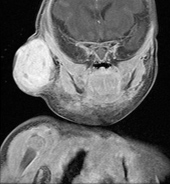
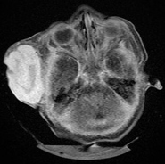
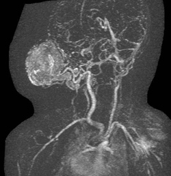
Beyond expert clinical evaluation, the use of radiological imaging is routinely used to better characterize the vascular anomaly and to delineate the extent of involvement. Among the studies we use are ultrasounds, CT (computerized tomography) scans, MRI (magnetic resonance imaging) scans, and MRA (magnetic resonance angiogram) scans.
 Diagnostic MRI (frontal) of large hemangioma |
 Diagnostic MRI (transverse) of large hemangioma |
|
 Diagnostic MRA of large hemangioma |
||
3-D image of large hemangioma: