Craniosynostosis is the premature closure or fusion of the open areas, or sutures, between the skull plates in an infant’s skull. When there is no other involvement besides the skull plates, the cause is usually unknown, and the condition is called non-syndromic craniosynostosis. When a suture closes prematurely, a predictable abnormality of head shape occurs due to compensatory expansion required by the growing brain. A skilled geneticist can determine if a child’s craniosynostosis is syndromic or non-syndromic.
Types of Non-Syndromic Craniosynostosis
 Metopic Craniosynostosis: The forehead portion of the skull becomes triangular in shape and the eyes become closer together (trigonocephaly).
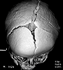
 Sagittal Craniosynostosis: The most common type of single suture fusion. The head becomes elongated and narrowed and takes on the shape of a boat, scaphocephaly.
 Coronal Craniosynostosis: When one coronal suture is fused, the orbit is pulled back and upward, while the opposite side grows down and forward to compensate. If both coronal sutures are involved, the entire forehead along with the orbital rims above the eyes are drawn backward (brachycephaly). Sometimes the head appears tall (turricephaly).
 Lambdoid Craniosynostosis: The head becomes trapezoidal in shape. This is the rarest of the craniosynostoses, accounting for only about 4 percent of cases.
Metopic Craniosynostosis
Metopic Craniosynostosis is one of the more common forms of this disorder, accounting for approximately 40 percent of all single-suture synostosis. The metopic suture lies along the midline of the forehead and, when fused prematurely, leads to a ridge in the middle of the forehead and a triangular shape to the skull (trigonocephaly). A small fraction of these patients will have increased intracranial pressure and other neurologic abnormalities. Correction is usually recommended for improvement in the cosmetic appearance, as well as to address any possible underlying neurologic concerns.
Metopic craniosynostosis is usually corrected for cosmetic reasons and an experienced craniofacial plastic surgeon will assure a strong focus on this objective.
Characteristics of metopic craniosynostosis include:
- Triangular shape of the anterior skull
- Triangular shape of the orbits
- Orbital hypotelorism
- Narrowing between the temples
Correction is generally carried out by a skilled pediatric craniofacial plastic surgeon and pediatric neurosurgeon team. The surgeons at Rady Children’s are among the most experienced craniofacial plastic surgeons in the world, ensuring a strong focus on the aesthetic outcome of corrective surgery. Our team is unparalleled in the management of these children and the use of the newest techniques and technologies.
Sagittal Craniosynostosis
Sagittal Craniosynostosis is one of the more common forms of this disorder, and like the metopic form, it accounts for approximately 40 percent of all single-suture synostosis. The sagittal suture lies along the midline of the skull. When this suture fuses prematurely, the head cannot grow in width, but must grow in length to accommodate the expanding brain.
Sagittal Craniosynostosis can be corrected within the first year of life using new endoscopic techniques and biodegradable technology.
Characteristics of sagittal craniosynostosis include:
- Elongated skull shape (front to back)
- Narrow skull shape (side to side)
- Midline bony ridge
- Frontal bossing
- Occipital prominence
When caught early, children with sagittal synostosis are candidates for a new minimally invasive endoscopic approach. The surgeons at Rady Children’s have developed a brand-new means of treating these children using a combination of endoscopy and immediate correction of the skull deformity with biodegradable plates and tacks, which are small rivets placed into the bone to stabilize the plates. This new approach reduces, and often completely eliminates, the need for the band or helmet molding of the skull that is used after traditional endoscopic approaches.
When children with sagittal synostosis present at older ages, correction involves cranial vault reconstruction, which can be carried out safely and simply using a standard coronal incision from ear to ear that is hidden in the hair. Whether corrected early or late, it is best performed by a skilled pediatric craniofacial plastic surgeon and pediatric neurosurgeon team.
Coronal Craniosynostosis
Coronal Craniosynostosis is a premature closure of the skull sutures that lie behind the forehead and run from side to side. Coronal craniosynostosis may be unilateral or bilateral. When both coronal sutures are involved, it is more likely that an underlying syndrome is present. Geneticists will determine if the condition is syndromic or non-syndromic.
The correction of coronal craniosynostosis using the new techniques possible with biodegradable technology permit a more precise achievement of the desired skull shape.
Characteristics of coronal craniosynostosis include:
- Flattening of one or both sides of the forehead
- Increased forehead height
- Widening of the skull (side to side)
- Recession of one or both brows
In children with bilateral coronal craniosynostosis, the pediatric craniofacial plastic surgeon and neurosurgeon work together as a team to correct the skull deformities and reposition the bones in their normal location, holding them there with biodegradable fixation devices that will gradually disappear over time. When children have unicoronal synostosis, a similar approach is preferred. Both sides are addressed, even when only one side is involved, because of the compensatory changes present in the opposite side. Rady Children’s craniofacial plastic surgeons have a vast experience with the correction of bilateral and unicoronal craniosynostosis.
Lambdoid Craniosynostosis
Lambdoid Craniosynostosis is quite rare and occurs in only 2 to 4 percent of patients with craniosynostosis. The lambdoid suture is located along the back of the head, and it may fuse prematurely on one side or on both sides. Typically, fusion will cause the skull to develop a trapezoid shape, indicating restricted growth at the fused suture and compensatory growth changes surrounding the suture.
Characteristics of lambdoid craniosynostosis include:
- Ridging over the fused lambdoidal suture
- Contralateral frontal bossing
- Posterior displacement of the ipsilateral ear
- Ipslateral occipitomastoid bulge
A diagnosis can usually be made by physical examination, but occasionally a CT scan is necessary to verify true fusion of the lambdoid suture. More commonly, a trapezoid-shaped head is the result of a positional head deformity, and the lambdoid suture is open. It is critical to differentiate the two disorders in the early months of life, as position-related head deformities are then easily treated by molding with bands or helmets rather than with surgical treatment. Craniofacial plastic surgeons work with geneticists and pediatric neurosurgeons to assure correct diagnosis and treatment.